
Evaluation of marginal adaptation and bond strength of apical root canal plugs using different bioceramic cements
Atualizado: 5 de ago.
DOI: https://doi.org/10.1186/s12903-026-07787-9
Journal: BMC Oral Health (2026) 26:332
Received: 22 January 2025 / Accepted: 22 January 2026 / Published online: 10 February 2026
Contact: Frank Ferreira Silveira — frankfoui@uol.com.br
How to cite: Lima MSFF, Antunes ANG, de Toubes KMPS, Bruzinga FFB, Caneschi CS, Morgan LFSA, Silveira FF. Evaluation of marginal adaptation and bond strength of apical root canal plugs using different bioceramic cements. BMC Oral Health. 2026;26:332.
Michel Sena Fernandes Faria Lima — Departamento de Odontologia, PUC Minas (¹)
Alberto Nogueira da Gama Antunes — Departamento de Odontologia, PUC Minas (¹)
Kênia Maria Pereira Soares de Toubes — Universidade de Uberaba (²)
Fábio Fernandes Borém Bruzinga — Departamento de Odontologia, PUC Minas (¹)
Camila de Sousa Caneschi — Faculdade de Odontologia, UFMG (³)
Luís Fernando dos Santos Alves Morgan — Faculdade de Odontologia, UFMG (³)
Frank Ferreira Silveira* — Departamento de Odontologia, PUC Minas (¹) — Corresponding author
¹ Departamento de Odontologia – Pontifícia Universidade Católica de Minas Gerais, Avenida Dom José Gaspar 500 / Prédio 46, Coração Eucarístico, Belo Horizonte, MG 30535-610, Brasil
² Universidade de Uberaba, Av. Nenê Sabino, 1801 - Universitário, Uberaba - MG 38055-500, Brasil
³ Faculdade de Odontologia – Universidade Federal de Minas Gerais, Rua Professor Moacir Gomes de Freitas 688, Pampulha, Belo Horizonte, MG 31270-901, Brasil
Competing interests: The authors declare no competing interests.
Funding: No funding was received for this research.
ABSTRACT
Background: Apexification in immature teeth represents a clinical challenge due to the difficulty of achieving an effective apical seal and sufficient mechanical resistance. Bioceramic materials such as MTA Repair HP, Bio-C Repair, Biodentine, and PBS Cimmo HP have been developed to improve sealing ability and bond strength. The objective of this study was to evaluate and compare, in vitro, the apical marginal adaptation by Scanning Electron Microscopy (SEM) and bond strength through the push-out test of these four calcium silicate-based cements used as an apical barrier.
Methods: Forty simulated models of immature teeth with open apex were created. The specimens were divided into four groups to create an apical plug: G1 (MTA Repair HP), G2 (Bio-C Repair), G3 (Biodentine) and G4 (Cimmo HP). The apical portion of the specimens was sectioned, into 2.5 mm slices. The adaptation of the apical barrier was assessed using SEM and the bond strength was evaluated by the push-out test. Data were analyzed by one-way ANOVA and Tukey's post hoc test (p<0.05).
Results: MTA Repair HP showed significantly better marginal adaptation compared to Cimmo HP (p<0.05), while no statistical difference were found among the other materials. In contrast, PBS Cimmo HP exhibited the highest bond strength, significantly superior to Bio-C Repair (p<0.05). No significant differences were found among the other materials. Both MTA Repair HP and Biodentine demonstrated consistent performance in both tests. Adhesive fracture was the most frequent type of failure observed across all materials.
Conclusion: MTA Repair HP, provided superior marginal adaptation, favoring its indication for apical sealing and prevention of leakage. Conversely, Cimmo HP, with its higher compressive strength, may be better suited for cases requiring greater mechanical resistance. Understanding the distinct properties of these materials assists clinicians in selecting the most appropriate bioceramic cement for apexification procedures.
Keywords: Endodontics; Bioceramic cements; Marginal adaptation; Calcium silicate; Apexification.
BACKGROUND
Endodontic treatment of teeth with incomplete root formation constitutes a clinical challenge for the dentist [1]. In cases like this, apexification can be performed in order to seal the apical foramen and induce the formation of a mineralized tissue [2, 3]. MTA Repair HP, Bio-C Repair, Biodentine and PBS Cimmo HP are calcium silicate-based cements used for this purpose [4–6].
Calcium hydroxide was the first material used to induce the formation of an apical barrier in teeth with an open apex, however its limitations include long treatment time, multiple clinical sessions and unpredictability results [1]. The mineral trioxide aggregate (MTA) emerged as a favorable alternative because it has qualities similar to those of calcium hydroxide and allows treatments to be carried out in a single session. Despite its advantages, conventional MTA presents handling difficulties and can cause coronal discoloration due to bismuth oxide, its radiopacifying agent [3, 7, 8].
To overcome these drawbacks MTA Repair HP was introduced, which introduced the change of radiopacifying agent to calcium tungstate and the addition of polyvinylpyrrolidone to distilled water to allow better handling [4, 8, 9]. Bio-C Repair is a new pre-manipulated option, facilitating use and speeding up application to the cavity [5, 10].
Biodentine, another calcium silicate-based cement, has been successfully used for dentin replacement and in procedures such as root perforation, apexification and regenerative endodontics treatments [7, 10]. PBS Cimmo HP® is a recently introduced calcium silicate-based cement. It contains pozzolan and calcium carbonate in order to increase its compressive strength. It is recommended for various endodontic procedures and has shown promising results in previous studies, demonstrating good biocompatibility and the formation of mineralized tissue surrounding the material [6, 11].
One of the remarkable characteristics of bioceramic materials is its ability to release calcium hydroxide upon contact with tissue fluids. This calcium hydroxide interacts with tissue phosphates, facilitating hydroxyapatite formation and inducing tissue regeneration [3, 9]. Notably, the crystallization of this material within the dentinal tubules creates a robust interface with the dentin. It contributes to enhanced resistance against microinfiltration, improved mechanical strength, and increased resistance to adhesion [9].
Although MTA is widely used, new materials with improved properties have been developed. In this sense, this study evaluated these new materials in comparison with MTA to determine whether they offer significant advantages in terms of marginal adaptation and bond strength. Several microleakage methods have been evaluated in previous studies to verify the marginal adaptation of apical barriers in immature teeth. Scanning Electron Microscope (SEM) has been used frequently as it allows evaluation through images and observation of defects in the desired magnification [12, 13]. The push-out test has been widely accepted to evaluate the bond strength of filling materials to dental tissue, as it is performed under conditions similar to clinical conditions [10].
The present study aimed to compare the marginal adaptation and bond strength of MTA Repair HP, Bio-C Repair, Biodentine, and PBS Cimmo HP, used as apical barriers in simulated immature teeth. Marginal adaptation was assessed by scanning electron microscopy (SEM), while bond strength was evaluated using the push-out test. The null hypothesis was that there would be no significant differences in marginal adaptation and bond strength among the tested materials.
METHODS
Sample selection
Extracted single-rooted human teeth, all exhibiting complete root formation and free of fracture lines, with an average length ranging from 22 to 25 mm, were selected from the tooth bank at the Pontifical Catholic University of Minas Gerais.
Additionally, they were submitted to ortho and mesioradial radiographs, and teeth with previous endodontic treatment, calcified root canals, abrupt curvature, or internal root resorption were excluded, resulting in a total of forty teeth. After selection, they were stored in a 0.1% thymol solution.
Sample Preparation
To simulate teeth with incomplete root formation, experimental models were created [14, 15]. Using a carborundum disc (SS White Artigos Dentários, Rio de Janeiro, Brazil), the dental crown was sectioned at the cementoenamel junction and the apical portion, to standardize the length at 10 mm, eliminating apical deltas and standardizing the output of the canal in the center of the root.
The root canals were prepared using Gates Glidden #6 to #1 drills (Maillefer, Ballaigues, Switzerland), in the crown-apex direction, with the #1 drill being used until it surpassed the apical foramen. At each instrument change, the canals were irrigated with 5.25% sodium hypochlorite solution (Lenza Farmacêutica, Dental Division, Belo Horizonte, Brazil).
A divergence in the apical preparation was performed through retrograde instrumentation using a Reciproc Blue #40 taper 0.06 file (VDW, Munich, Germany), inserted along the entire length of its active part, marked with a rubber cursor. After final irrigation with 17% EDTA solution (Biodinâmica Química e Farmacêutica, Ibiporã, PR, Brazil) for 5 min, the canals were dried with #80 absorbent paper points (MK Life, Porto Alegre, Brazil), and the 40 apical foramina were microscopically evaluated for shape and integrity at 13x magnification (M-900 DF Vasconcelos, São Paulo, Brazil). Specimens were wrapped in damp gauze and maintained in an incubator at 37 °C (NL-80-27, New Lab, Piracicaba, Brazil) to prevent desiccation.
Construction of the apical barrier
The samples were randomly divided into four groups: G1: MTA Repair HP (n = 10); G2: Bio-C Repair (n = 10); G3: Biodentine (n = 10) and G4: PBS Cimmo HP (n = 10) (Table 1). The sample size was determined based on a previous study [14, 15]. The handling and insertion of each material into the root canal were carried out according to the manufacturer's recommendations, assisted by an MTA applicator (Angelus, Londrina, Brazil). A wet foam attached to a bench vice was used to simulate the periapical tissues and offer resistance to the extrusion of the material when creating the apical plug.
The material was compacted using Schilder condensers (Endoprime, Santa Luzia, Brazil) to a depth of 7 mm, followed by indirect ultrasonic vibration (ENAC, Osada Incorporated, Los Angeles, USA) for 5 s, without irrigation. The width and quality of the apical barriers were checked by ortho and mesio-radial radiographs. The roots were kept in damp gauze and maintained in an incubator at 37 °C for thirty days until the molds were taken.
Table 1. Information on bioceramic cements. Composition described according to the leaflet.

Molding using the replica technique
The apical surface of each root was inspected and polished with abrasive silicon carbide sandpaper of grain size 800, 1500 and 2000, respectively, for 10 s, under refrigeration and water. The roots were washed under running water and dried with an air jet after polishing.
To obtain negative replicas, the specimens were molded using fluid paste addition silicone (Futura AD, DFL Indústria e Comércio S/A, Rio de Janeiro, Brazil). After setting, positive replicas were obtained by pouring G4 rigid epoxy resin (Polipox, Cesário Lange, Brazil) onto the molds, manipulated following the manufacturer's instructions. After taking prey, the replicas were separated.
Scanning electron microscope (SEM) analysis
For SEM analysis (JSM-6510LV, JEOL, Tokyo, Japan), the replicas were blasted with an air jet and fixed in aluminum stubs with the apical portion facing upwards. They were then taken to the metalizer (Desk V, Denton Vacuum, Moorestown, USA).
After this process, the replicas were analyzed and photographed with the SEM, capturing an image at 60x magnification to observe the apical foramen region. Four points were marked on each replicate (Fig. 1) for image capture at 700x magnification. The images were saved in TIFF format using the SEM Control User Interface software – Version 2.01 (JEOL Technics, Japan) at a resolution of 1280 × 1024 pixels.
Fig. 1 SEM image of an apex at 60x magnification. Image corresponding from a G2 specimen. Numbers in yellow represent the points selected for evaluation at 700x magnification.

With the aid of Image J software (National Institute of Health, Bethesda, USA), each image obtained at 700x magnification was evaluated separately, using the scale present in the photomicrographs as a calibration method. The measurement of the total area corresponding to the misadaptation was carried out using the polygon tool, also recording the gap with the largest and smallest area in square micrometers (µm²) (Fig. 2).
Fig. 2 SEM image showing the measurement of the area of different gaps at 700x magnification. Image corresponding to point 3 of a G3 specimen, showing the measurement of the area of different gaps, with the largest gap area being 664.61 µm², the smallest gap area 39.77 µm² and the total area corresponding to the sum of all areas, amounting to 877.96 µm².

Furthermore, using the line tool, the total measurement of the dentin wall and the measurement of the dentin wall with a gap were obtained, in micrometers (µm), to determine the percentage of the dentin wall with marginal misadaptation (Fig. 3). The largest and smallest gaps were also measured, in micrometers (µm), perpendicular to the dentin wall (Fig. 4).
Fig. 4 SEM image showing the length of the gaps at 700x magnification. Image corresponding to point 1 of a G4 specimen, showing the length of the gaps between the dentin wall (top) and the material (bottom), with a measurement of 26.28 µm for the largest gap and 4.14 µm for the smallest gap.

Fig. 3 SEM image showing a % dentin misadaptation of the material at 700x magnification. Image corresponding to point 3 of a G4 specimen, showing a % dentin misadaptation of the material of 28.35%, obtained through the ratio between the length of the dentin wall with gap and the total length of the wall dentin in the image. A: Total length of the dentin wall, measuring 143.20 µm. B: Length of the dentin wall with a gap, measuring 40.60 µm.

Cutting of specimens
Each specimen was fixed on acrylic resin plates using godiva (Godiva Exata, DFL Indústria e Comércio S/A, Rio de Janeiro, Brazil) and keeping its long axis parallel to the long axis of the plate. The resin plates were coupled to a precision cutting machine (IsoMet™ 1000, Buehler, Lake Bluff, USA) and, using a 0.5 mm thick diamond disc (IsoMet™ Diamond Wafering Blades, Buehler, Lake Bluff, USA), under constant refrigeration at a speed of 400 rpm, a section was made in the mesiodistal direction, perpendicular to the long axis of each specimen, obtaining a 2.5 mm thick slice.
Push-out test
The slices were positioned individually on a stainless-steel metal base (Instron 3344, Instron, Canton, USA) that has a 2.5 mm diameter hole. The apical portion of the slice was facing downwards, in contact with the metal base.
A metal rod with an active tip measuring 0.8 mm in diameter was used, attached to the upper portion of the universal testing machine (EZ-LX, Shimadzu, Kyoto, Japan) and positioned over the apical plug. The test was carried out with a 5 kN load cell, at a speed of 0.5 mm/min in the coronal/apex direction, until the maximum force for failure occurred (Fig. 5).
Fig. 5 Push-out bond strength test. Illustration of the methodological procedure.

Once the push-out was complete, the area of the adhesion surface was calculated, in mm², using the formula: A = π (R2 + R1) [h² + (R2 - R1)²]⁰·⁵, in which π = 3.14, R2 = apical radius of the plug (larger radius), R1 = coronal radius of the plug (smaller radius) and h = thickness of the slice. Then, to obtain the adhesive strength values in MPa, the ratio between the maximum force exerted (N) and the area of the adhesion surface was calculated [16].
After the push-out test, the slices were collected and microscopically inspected under a surgical microscope at 13x magnification (M-900 DF Vasconcelos, São Paulo, Brazil) to visualize the type of fracture obtained. Failures were classified as adhesive (rupture of the bond at the dentin/material interface), cohesive (rupture within the material), and mixed (adhesive rupture in the dentin and cohesive rupture within the material).
Statistical analysis
Initially, the data were subjected to the Kolmogorov-Smirnov normality test, which demonstrated normal distribution in all variables. Then, the mean and standard deviation were calculated for each specimen and, after that, for the group as a whole.
To evaluate the differences between the variables in the four groups, the one-factor Analysis of Variance test was used followed by Tukey's post hoc test (p > 0.05). The analysis was carried out separately for the bond strength and for each variable related to the gap present. The significance level was set at 5%. Analyzes were performed using the BioEstat 5.3 software (BioEstat, Belém, Brazil).
RESULTS
Analysis of SEM photomicrographs showed superior results from MTA Repair HP (G1) in relation to Cimmo HP (G4). There was no difference between the groups in the variables % gap per µm of dentin and smallest gap area (µm²). There was a statistically significant difference between MTA Repair HP (G1) and Cimmo HP (G4), in the variables total gap area (µm²) (p < 0.01), largest gap (µm) (p < 0.05), smallest gap (µm) (p < 0.05), smallest gap area (µm²) (p < 0.01). Bio-C Repair (G2) and Biodentine (G3) did not show a significant difference between the other groups (Table 2).
The push-out test resulted in better results for Cimmo HP (G4) in relation to Bio-C Repair (G2), showing a statistically significant difference (p < 0.05). MTA Repair HP (G1) and Biodentine (G3) did not show a significant difference between the other groups (Table 3).
MTA Repair HP showed excellent results in both tests, with values similar to Biodentine and Bio-C Repair. Cimmo HP presented the best result in terms of bond strength in comparison to Bio-C Repair, although without difference of Biodentine and MTA Repair HP.
The results of the type of fracture obtained were represented as a percentage (Table 4). Overall, the groups followed the same pattern. While MTA Repair HP presented fewer adhesive fractures than the others (70%), it was the material that presented the most mixed fractures (20%). On the other hand, Bio-C Repair presented the most adhesive fractures (90%) and did not present mixed fractures. All groups presented 10% cohesive fracture.
Table 2. Mean and standard deviation of marginal adaptation variables and comparison between groups.
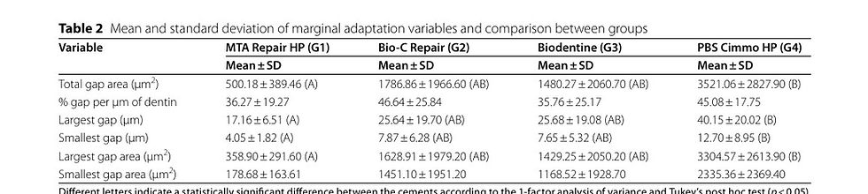
Different letters indicate a statistically significant difference between the cements according to the 1-factor analysis of variance and Tukey's post hoc test (p < 0.05).
Table 3. Mean and standard deviation of bond strength and comparison between groups.

Mean and standard deviation of the push-out test. 1-factor analysis of variance and Tukey post-hoc test with a significance level of 95%. Different letters indicate statistically significant differences.
Table 4. Frequency (%) of fracture types in each group.

-: Absence of type of fracture.
DISCUSSION
Although conventional MTA has wide clinical application and is considered the material of choice for treatments such as creating an apical barrier, new materials have been developed with better properties and free from impurities [10].
Materials such as MTA Repair HP and Biodentine present improvements in their physical properties related to manipulation [17, 18]; and Bio-C Repair is sold in pre-manipulated format, maintaining the favorable properties of conventional MTA [19]. Another improvement was the change in the radiopacifying agent, no longer causing staining of the coronary structure [8].
Lack of adaptation between the cement and dentin can lead to apical infiltration, leading to periapical changes and possibly treatment failure. Therefore, good adaptation and adhesion of the filling material to the root dentin are essential requirements [12, 14].
Several methods for evaluating microleakage have been evaluated in previous studies, but SEM has been used to evaluate gaps in studies that aim to evaluate the marginal adaptation of apical barriers in immature teeth [13, 20]. Although SEM is a suitable method for evaluating the surface topography of the sample, it is not possible to perform evaluations in three dimensions [21].
Gondim Jr et al. [22] showed that the details observed in a hard tissue analysis did not differ from the images taken, both in the original tooth and in the resin replicas. However, SEM evaluation using the original tooth specimen may present complications such as cracks in the hard tissue of the samples due to dehydration and movement or separation of the filling material from the dentin walls [23, 24].
Gondim Jr et al. [25] evaluated the marginal adaptation of the completion of the filling in terms of gap area, using magnifications of 150x and 300x across the entire apical foramen. The measurement of the gap area is relevant because it is associated with apical infiltration, and can be analyzed through 3D profilometry, which has the ability to measure the maximum depth formed of the gaps [14, 26, 27].
The gap percentage was also used by Aguiar et al. [28], relating the perimeter of the dentin wall with material misadaptation to the total perimeter. Furthermore, the width and length of the largest and smallest gaps are relevant measurements, having been used in studies to evaluate the apical marginal adaptation of different materials [12, 14, 15, 21, 23].
To avoid distortions in the results, points 1, 2, 3 and 4 of all specimens followed a standardized protocol to capture images at higher magnification, totaling 40 points per group, in accordance with what was done by Badr [29]. Other studies evaluated different numbers of areas per specimen or per group, including 4 areas per tooth for a total of 80 per group, 8 areas at each apex for a total of 576 areas evaluated, and 12 areas per specimen for a total of 18 teeth [15, 21, 23].
In the current study, there was no difference between the groups in terms of gap percentage. In contradiction to this result, Aguiar et al. [28] observed worse results with MTA Repair HP in terms of gap percentage when compared with MTA Angelus and Biodentine (p < 0.05). In another study, Alazrag et al. [30] obtained favorable results with Biodentine when compared to TheraCal LC and MTA Angelus (p < 0.05).
In this study, there was a statistical difference in the gap length only between MTA Repair HP and Cimmo HP (p < 0.05), with no difference between Bio-C Repair and Biodentine. De Sá et al. [14] found a similar result. In a similar study, Soundappan et al. [31] evaluated the length of the gap between MTA, Biodentine and IRM, obtaining better results for MTA and IRM when compared with Biodentine (p < 0.05). The presence of pozzolan in Cimmo HP may have negatively influenced the results in this study by reducing its setting time [6].
The push-out test has been widely used to evaluate the bond strength of filling materials to dentin tissue, but there is a lack of standardization, both in the test and in the specimen preparation method, which can result in discrepant results [10, 17, 19].
Nagas et al. [32] previously showed that the diameter of the active tip used in the push-out tests, which ranged from 50% to 83% of the channel diameter, did not affect the values obtained in the test. In this study, a device with a 0.8 mm active tip (66% of the canal diameter) was used.
In the current study, Cimmo HP performed better in the push-out test compared to Bio-C Repair (p < 0.05). There was no statistical difference between the other materials. This result may have occurred due to the addition of pozzolan and calcium carbonate to Cimmo HP, in order to increase the material's compressive strength [14]. Although Bio-C Repair is a pre-manipulated material, more studies are needed to evaluate its physicochemical properties in comparison with other materials.
In accordance with the current study, de Sá et al. [14] found no difference in the push-out test between MTA Angelus, MTA Repair HP and Biodentine. However, Rodrigues et al. [19] and Aguiar et al. [28], when comparing the materials MTA Angelus, MTA Repair HP, Biodentine and Bio-C Repair, they found a better result for Biodentine (p < 0.05).
This in vitro study has inherent limitations, such as not fully replicating the complex biological conditions of the oral environment. Additionally, variations in application techniques and the duration of evaluation may influence the results, limiting their direct clinical applicability. Thus, it is important to consider the limitations of the methodology used in relation to apical barrier construction and gap assessment. If we consider sealing ability as the reproduction of the seal in the entire canal perimeter, a gap may not represent what has actually occurred in the entire apical plug. Therefore, it is extremely difficult to establish a correlation between sealing ability and marginal adaptation. Additionally, the absence of simulated tissue pressure during material setting may have influenced the material's behavior compared to what occurs in clinical conditions.
Considering the importance of the clinical approach to be carried out in teeth with an open apex, the findings of the present study suggest that MTA Repair HP, given its improved properties, is an excellent material to be used as an apical barrier. Despite Bio-C Repair being pre-mixed for easier handling, it is necessary to carry out additional studies to evaluate its bond strength as an apical barrier. Biodentine, a material widely used in various treatments, showed excellent results. Cimmo HP, as it is a new material on the market with clinical applications similar to MTA, although it has shown good results in the push-out test, requires further studies to evaluate its marginal adaptation in immature teeth. More in vivo studies are needed to confirm these findings.
CONCLUSIONS
The results showed that MTA Repair HP, due to its superior marginal adaptation, is more suitable for achieving a better seal and preventing leakage. Conversely, Cimmo HP, with its higher compressive strength, may be better suited for cases requiring greater mechanical resistance. Understanding these properties helps clinicians select the most appropriate material for successful treatment of immature teeth or open apex cases.
References
1. Rafter M. Apexification: a review. Dent Traumatol. 2005;21(1):1–8.
2. Torabinejad M, Nosrat A, Verma P, Udochukwu O. Regenerative endodontic treatment or mineral trioxide aggregate apical plug in teeth with necrotic pulps and open apices: a systematic review and meta-analysis. J Endod. 2017;43(11):1806–20.
3. Shabahang S. Treatment options: apexogenesis and apexification. Pediatr Dent. 2013;35(2):125–8.
4. Galarça AD, Da Rosa WLO, Da Silva TM, Lima GS, Carreño NLV, Pereira TM, et al. Physical and biological properties of a High-Plasticity tricalcium silicate cement. Biomed Res Int. 2018;2018Nov27:8063262.
5. Ghilotti J, Sanz JL, López-García S, Guerrero-Gironés J, Pecci-Lloret MP, Lozano A, et al. Comparative surface Morphology, chemical Composition, and cytocompatibility of Bio-C Repair, Biodentine, and proroot MTA on hDPCs. Mater (Basel). 2020,13(9):2189.
6. Santiago MC, Gomes-Cornélio AL, de Oliveira LA, Tanomaru-Filho M, Salles LP. Calcium silicate-based cements cause environmental stiffness and show diverse potential to induce osteogenesis in human osteoblastic cells. Sci Rep. 2021;18(1):16784.
7. Coaguila-Llerena H, Ochoa-Rodriguez VM, Castro-Núñes GM, Faria G, Guerreiro-Tanomaru JM, Tanomaru-Filho M. Physicochemical properties of a bioceramic repair material – BioMTA. Braz Dent J. 2020;31(5):511–5.
8. Ferreira CMA, Sassone LM, Gonçalves AS, de Carvalho JJ, Tomás-Catalá CJ, García-Bernal D, et al. Physicochemical, cytotoxicity and in vivo biocompatibility of a high-plasticity calcium-silicate based material. Sci Rep. 2019;8(1):3933.
9. Pelepenko LE, Saavedra F, Antunes TBM, Bombarda GF, Gomes BPFA, Zaia AA et al. Investigation of a modified hydraulic calcium silicate-based material – Bio-C Pulpo. Braz Oral Res. 2021;35:e077. https://doi.org/10.1590/1807-3107br-2021.vol35.0077. PMID: 34161414.
10. Lucas CPTR, Viapiana R, Bosso-Martelo R, Guerreiro-Tanomaru JM, Camilleri J, Tanomaru-Filho M. Physicochemical properties and dentin bond strength of a tricalcium silicate-based retrograde material. Braz Dent J. 2017;28(1):51–6.
11. Pedrosa MDS, Vilela HDS, Rahhal JG, Bueno NP, Lima FS, Nogueira FN, et al. Response of periodontal ligament stem cells to lipopolysaccharide and calcium silicate-based materials. Braz Dent J. 2022;33(2):73–82.
12. Polineni S, Bolla N, Mandava P, Vemuri S, Mallela M, Gandham VM. Marginal adaptation of newer root canal sealers to dentin: a SEM study. J Conserv Dent. 2016;19(4):360–3.
13. Khalighinejad N, Barekatne B, Hasheminia SM, Gheibolahi H. In vitro evaluation of two methods of ultrasonic irrigation on marginal adaptation of MTA plugs in open apex teeth: a SEM analysis. Indian J Dent Res. 2014;25(1):69–72.
14. de Sá MAB, Nunes E, Antunes ANG, Brito Júnior M, Horta MCR, Amaral RR, et al. Push-out bond strength and marginal adaptation of apical plugs with bioactive endodontic cements in simulated immature teeth. Restor Dent Endod. 2021;46(4):e53.
15. Araújo AC, Nunes E, Fonseca AA, Cortes MI, Horta MC, Silveira FF. Influence of smear layer removal and application mode of MTA on the marginal adaptation in immature teeth: a SEM analysis. Dent Traumatol. 2013;29(3):212–7.
16. Goracci C, Sadek FT, Monticelli F, Cardoso PEC, Ferrari M. Microtensile bond strength of self-etching adhesives to enamel and dentin. J Adhes Dent. 2004;6(4):313–8.
17. Akbulut MB, Bozkurt DA, Terlemez A, Akman A. The push-out bond strength of biofactor mineral trioxide aggregate, a novel root repair material. Restor Dent Endod. 2019;44(1):e5.
18. Guimarães BM, Prati C, Duarte MAH, Bramante CM, Gandolfi MG. Physicochemical properties of calcium silicate-based formulations MTA repair HP and MTA Vitalcem. J Appl Oral Sci. 2018;26:e2017115. https://doi.org/10.1590/1678-7757-2017-0115. PMID: 29641748; PMCID: PMC5912399.
19. Rodrigues MNM, Bruno KF, de Alencar AHG, Silva JDS, de Siqueira PC, Decurcio DA, et al. Comparative analysis of bond strength to root dentin and compression of bioceramic cements used in regenerative endodontic procedures. Restor Dent Endod. 2021;46(4):e59.
20. Zafar M, Iravani M, Eghbal MJ, Asgary S. Coronal and apical sealing ability of a new endodontic cement. Iran Endod J. 2009;4(1):15–9.
21. Bidar M, Moradi S, Jafarzadeh H, Bidadi S. Comparative SEM study of the marginal adaptation of white and grey MTA and Portland cement. Aust Endod J. 2007;33(1):2–6.
22. Gondim E Jr, de Gomes BPFA, Ferraz CCR, Teixeira FB, de Souza-Filho FJ. Investigation of the marginal adaptation of root-end filling materials in root-end cavities prepared with ultrasonic tips. Int Endod J. 2003;36(7):491–9.
23. Ghorbanzadeh A, Shokouhinejad N, Fathi B, Raoof M, Khoshkhounejad M. An in vitro comparison of marginal adaptation of MTA and MTA-like materials in the presence of PBS at one-week and two-month intervals. J Dent (Tehran). 2014;11(5):560–8.
24. Torabinejad M, Smith PW, Kettering JD, Pitt Ford TR. Comparative investigation of marginal adaptation of mineral trioxide aggregate and other commonly used root-end filling materials. J Endod. 1995;21(6):295–9.
25. Gondim E Jr, Zaia AA, Gomes BPFA, Ferraz CCR, Teixeira FB, Souza-Filho FJ. Investigation of the marginal adaptation of root-end filling materials in root-end cavities prepared with ultrasonic tips. Int Endod J. 2003;36(7):491–9.
26. Gundam S, Patil J, Venigalla BS, Yadanaparti S, Maddu R, Gurram SR. Comparison of marginal adaptation of mineral trioxide aggregate, glass ionomer cement and intermediate restorative material as root-end filling materials, using scanning electron microscope: an in vitro study. J Conserv Dent. 2014;17(6):566–70.
27. Munhoz MF, Marchesan MA, Cardoso DRF, Silva SCR, Silva-Sousa YTC, Sousa-Neto MD. Quantitative 3D profilometry and SEM analysis of the adaptation of root-end filling materials placed under an optical microscope. Int Endod J. 2011;44(6):510–6.
28. Aguiar BA, Frota LMA, Taguatinga DT, Vivan RR, Camilleri J, Duarte MAH, et al. Influence of ultrasonic agitation on bond strength, marginal adaptation, and tooth discoloration provided by three coronary barrier endodontic materials. Clin Oral Investig. 2019;23(11):4113–22.
29. Badr AE. Marginal adaptation and cytotoxicity of bone cement compared with amalgam and mineral trioxide aggregate as root-end filling materials. J Endod. 2010;36(6):1056–60.
30. Alazrag MA, Abu-Seida AM, El-Batouty KM, El Ashry SH. Marginal adaptation, solubility and biocompatibility of theracal LC compared with MTA-angelus and Biodentin as a furcation perforation repair material. BMC Oral Health. 2020;20(1):298.
31. Soundappan S, Sundaramurthy JL, Raghu S, Natanasabapathy V. Biodentine versus mineral trioxide aggregate: randomized controlled trial. J Dent (Tehran). 2014;11(2):143–9.
32. Nagas E, Uyanik O, Durmaz V, Cehreli ZC. Effect of plunger diameter on the push-out bond values of different root filling materials. Int Endod J. 2011;44(10):950–5.
Authors' contributions
MSFL wrote the project, implemented the study methodology, contributed to the evaluation of the results, and wrote the manuscript.
ANGA contributed to the development of the research methodology, participated in the statistical analysis, and was involved in the manuscript revision.
KMPST contributed to the project design and played a significant role in the manuscript revision.
FFBB performed the statistical analysis of the collected data and contributed to the evaluation of the results.
CSC contributed to the laboratory work of the research conducted at the Federal University of Minas Gerais.
LFSAM contributed to the laboratory work of the research conducted at the Federal University of Minas Gerais.
FFS supervised the research. He conceptualized the methodology, participated in the laboratory work, and played a significant role in the manuscript revision.


Comentários